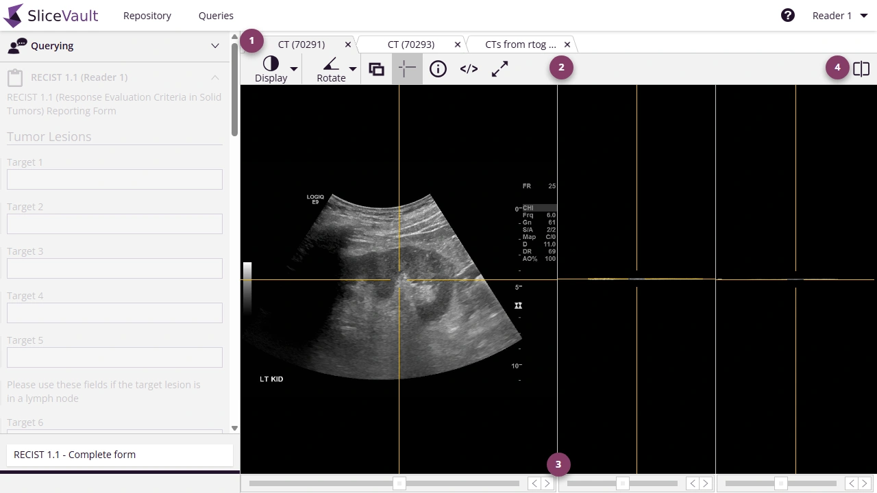
DICOM Viewer
SliceVault includes a built-in DICOM viewer that allows users to review medical images directly in the browser without requiring external software. The viewer provides essential tools to support quality control and central reading activities, while ensuring images remain securely stored in SliceVault.
Note: The SliceVault DICOM viewer is intended for use within the study workflow. It is not a replacement for full-featured diagnostic PACS systems. The embedded DICOM viewer is not CE marked or approved as a diagnostic medical device. It is provided solely for research and study workflow purposes.
-
Change Image – View all images uploaded for the visit, switch between tabs, and close image tabs as needed.
-
Display Controls – Use the display control bar to adjust visualization settings. See Viewer Controls below for details.
-
Change Slice – Navigate through image slices using the toolbar below the image.
-
Change View – Adjust the default layout to display multiple images side by side.
Viewer Controls
| Icon | Description |
|---|---|
 | Access display tools, including windowing, Gauss smoothing, interpolation tools, and more. |
 | Start/stop and change speed for images with timeseries. |
 | Switch plane: multi-planar reconstruction, transaxial plane only, transverse plane only, or sagittal plane only. |
 | Show DICOM tags. |
 | Reset zoom to default. |
 | Measure distance/angle between points. |
 | Show patient information in the viewer. |
 | Change default view, e.g., show multiple images side by side. |
Display Menu
The display menu lets you adjust how the active image is shown on screen without changing the uploaded data itself.
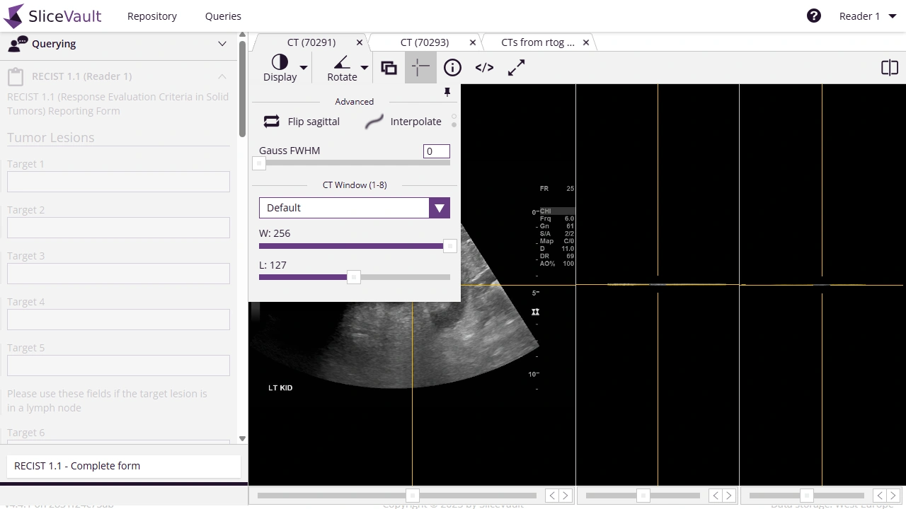
The exact options depend on the image type and study configuration, but the display menu is typically used for:
- Changing windowing or brightness/contrast presentation
- Applying smoothing or interpolation options
- Adjusting how PET or other overlay data is rendered
- Switching between available display presets for the active image
These tools are most useful when the image is hard to interpret with the default presentation, for example if soft tissue contrast is difficult to see or PET/CT overlays need a different rendering.
Hotkeys
Basic Viewing Tools
- Left mouse + drag – Move image
- Right mouse + drag up/down – Zoom out/in
- R – Reset zoom
- W / Scroll wheel up / ↑ – Move to previous slice
- S / Scroll wheel down / ↓ – Move to next slice
- C – Switch between available images
- V – Switch between available planes
- F – Enter full screen
- Esc – Exit full screen
- T – Triangulate
- M – Measure distance between two points
Viewing Tools for CT and Hybrid Imaging (PET/CT and SPECT/CT)
- 1–7 – Change CT window preset
- Middle click + drag up/down – Adjust CT window length manually
- Middle click + drag left/right – Adjust CT window width manually
- Ctrl + Alt + F – Flip sagittal image
- P – Toggle opacity between PET/SPECT and CT
- O – Toggle PET/SPECT smoothing (none, medium, high)
- N – Measure SUVmax in a circular ROI
Viewing Tools for Managing Images
- Shift + Left Click (on close tab) – Close all tabs with the same image type or series description within the active visit.
- Ctrl + Shift + Left Click (on close tab) – Close all tabs with the same image type or series description across all open visits.
- Ctrl + Left Click (on close tab) – Close all tabs with the same image ID across all open visits.
For image rendering problems, see Troubleshooting.